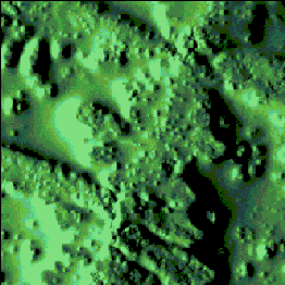
Virus particles on HOPG.
Scan size 1,1 micron
Poliomyelitis virus. AFM study.
 |
Virus particles on HOPG. Scan size 1,1 micron |
Virus particles on the NaCl crystal (substrate is mica). Scan size 5,8 micron |
 |
Conference:
Probe microscopy - 98
Nyzny Novgorod, 2-5
March
Authors:
Yaminsky I.V. , Bolshakova A.V.,
Faculty of Physics, Moscow
State University, Moscow
Loginov B.A.,
Protasenko V.V.,
ZAO "KPD",
Zelenograd
Suvorov A.L., Kozodaev
M.A., Volnin D.S.
Instituite of theoretical
and experimental physics, Moscow
Abstract:
Poliomyelitis virus
investigation using scanning tunneling and atomic force microscope is performed. Virus
particles are immobilized on the surface of different substrates (mica, HOPG, gold,
silver, titanium oxide). The sensitivity of scanning probe microscopy methods to detect
virus particles is estimated.
Back to the main page